Regenerative Medicine
Regenerative Medicine: The Power to Heal Your Body From Within.
Imagine a future where your body’s own biological toolkit is used to repair injury, rejuvenate tissues, and combat the effects of aging. This is not science fiction. This is the reality of regenerative medicine, a revolutionary field that is shifting the paradigm of healthcare from managing symptoms to promoting true, intrinsic healing. Instead of relying solely on external drugs or surgery to patch up damage, regenerative therapies harness and amplify the body’s natural repair mechanisms. By utilizing potent biological components like stem cells, growth factors from your own blood, and powerful cellular messengers, physicians can now target the root cause of an ailment. They can work to rebuild damaged cartilage, reduce chronic inflammation, and restore function to tissues that were previously considered beyond repair. This is medicine that works with your body, not just on it, unlocking a powerful new chapter in human health and longevity.
A Clinicians Guide To The Different Types Of Stem Cells
Understanding the different types of stem cells is fundamental for any clinician entering the regenerative space. Stem cells are broadly categorized by their potential to differentiate into other cell types. Embryonic stem cells (ESCs), which are a major focus for pioneering research institutions, are pluripotent meaning they can become any cell type in the body. While they hold immense therapeutic promise their use is fraught with ethical controversy and regulatory hurdles. Adult stem cells on the other hand are found in various tissues throughout the body and are generally multipotent. This means they can differentiate into a limited range of cell types usually specific to their tissue of origin.
The most commonly utilized adult stem cells in clinical practice are hematopoietic stem cells (HSCs) found in bone marrow and mesenchymal stem cells (MSCs). HSCs are the basis for bone marrow transplants and are crucial for treating blood-related disorders. MSCs are the workhorses of orthopedic and aesthetic regenerative medicine. They can be sourced from bone marrow, adipose (fat) tissue, and perinatal tissues like umbilical cord tissue. These cells are not just valuable for their ability to become bone, cartilage, and fat cells but primarily for their powerful signaling capabilities.
The Mechanism Of Action Of Mesenchymal Stem Cells
The true power of mesenchymal stem cells (MSCs) lies less in their ability to become new tissue and more in their role as masterful conductors of the healing process. Their primary mechanism of action is through paracrine signaling. This means they release a host of bioactive molecules including growth factors, cytokines, and extracellular vesicles like exosomes into their local environment. These signals create a pro-healing and anti-inflammatory microenvironment that orchestrates the body’s own repair crews.
When introduced to an area of injury or degeneration MSCs act as intelligent drugstores releasing specific factors in response to the environmental cues they receive. They can suppress local inflammation which is a major driver of pain and tissue damage. They can prevent existing cells from undergoing apoptosis or programmed cell death. Furthermore they can stimulate local progenitor cells to activate and begin the repair process and they can promote the formation of new blood vessels a process called angiogenesis which is critical for delivering oxygen and nutrients to healing tissue.
What Are Very Small Embryonic Like Stem Cells Vsels
A fascinating and still somewhat controversial area of stem cell research involves Very Small Embryonic-Like Stem Cells or VSELs. These are a rare population of cells found in adult tissues including bone marrow and peripheral blood. They are unique because they are very small and share certain markers with pluripotent embryonic stem cells. Proponents suggest that VSELs are a quiescent or dormant population of pluripotent stem cells residing within our bodies serving as a reserve for tissue repair and regeneration throughout life.
The theory is that under conditions of severe stress or injury these VSELs can be activated. Once activated they can differentiate into cells of all three germ layers ectoderm, mesoderm, and endoderm. This gives them a theoretical potential far beyond that of typical multipotent adult stem cells. While the research is compelling the scientific community is still working to fully characterize these cells and establish standardized protocols for their isolation and clinical use. Their potential represents a significant frontier in understanding the body’s innate regenerative capacity.

The Science Of Cell Signaling For Physicians
For physicians practicing regenerative medicine, mastering the language cells use to communicate is more critical than memorizing specific cell types. Healing is not about simply injecting cells it is about initiating a precise biological conversation. Cell signaling is the language cells use to communicate. This communication dictates everything from reducing inflammation to building new tissue. The primary mode of action for most regenerative biologics is the paracrine effect where cells release molecules that act on nearby cells.
This molecular dialogue involves a complex soup of growth factors, cytokines, and chemokines. Growth factors like Platelet-Derived Growth Factor (PDGF) and Transforming Growth Factor-beta (TGF-β) stimulate cell proliferation and matrix formation. Cytokines like Interleukin-1 Receptor Antagonist (IL-1RA) are powerfully anti-inflammatory blocking the destructive pathways common in conditions like osteoarthritis. Understanding this signaling cascade allows a clinician to choose the right biologic for the right condition and to explain to patients that the therapy is not just replacing tissue but is actually recruiting and directing the body’s own healing response.
A Comparison Of Prp Mscs And Exosomes For Orthopedics
In the orthopedic setting, clinicians must understand the nuances of several powerful tools, as each biologic has its own unique characteristics and applications. Platelet-Rich Plasma (PRP), Mesenchymal Stem Cells (MSCs), and Exosomes form the core of modern orthopedic biologics. PRP is the most accessible option derived from the patient’s own blood. It is a concentrate of platelets which release a robust cocktail of growth factors that can kickstart the healing cascade and reduce inflammation. It is often considered a first-line regenerative therapy for mild to moderate conditions.
MSCs sourced from bone marrow or adipose tissue offer a more powerful and sophisticated therapeutic action. They are living cells that not only release growth factors like PRP but also act as intelligent modulators of the joint environment for a sustained period. They are particularly effective at orchestrating a complex anti-inflammatory and regenerative response making them suitable for more significant injuries or degenerative conditions. Exosomes represent the next frontier. These are tiny vesicles released by MSCs containing their signaling cargo like mRNA and microRNA. They are the communication part of the MSC without the cell itself offering a potent anti-inflammatory and regenerative signal without the complexities of administering live cells.

Protocols For Preparing And Administering Platelet Rich Plasma Prp
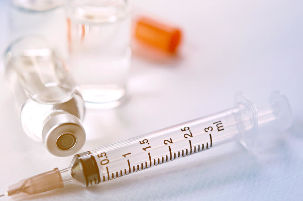
Because there is no single “PRP” but rather a spectrum of different formulations, the efficacy of Platelet-Rich Plasma (PRP) therapy is highly dependent on the specific methods used for its preparation and administration. The process begins with a standard blood draw from the patient. This blood is then placed into a centrifuge a machine that spins at high speeds to separate the blood components based on their density. The goal is to separate the platelets and a small volume of plasma from the red blood cells and the bulk of the plasma.
Key variables in preparation protocols include the centrifugation speed and time the use of a single-spin or dual-spin system and the decision to include or exclude the white blood cells (leukocytes). Leukocyte-rich PRP may be beneficial in certain tendon injuries but can cause a greater inflammatory flare in an arthritic joint. Therefore leukocyte-poor PRP is often preferred for intra-articular injections. Once prepared the PRP is carefully drawn into a syringe and administered to the target tissue often under ultrasound guidance to ensure precise placement.

Vetting Centrifuges And Preparation Systems For Prp
Not all PRP is created equal, and the centrifuge and preparation system used are the most critical factors in determining the quality of the final product. When vetting a system, clinicians must look beyond marketing claims and ask for specific performance data to ensure the final product is truly therapeutic. A high-quality system should provide verifiable data on its platelet concentration factors. A 3x to 5x concentration over baseline blood levels is generally considered the minimum for a therapeutic product.
Clinicians should also evaluate the system’s flexibility. Can it produce both leukocyte-rich and leukocyte-poor PRP? This versatility allows the treatment to be tailored to the specific patient and condition. Other important considerations include the cost per kit the sterility of the disposable components and the ease of use. Reputable companies will provide clinicians with scientific literature and validation studies for their systems. Choosing a validated high-quality system is a cornerstone of providing responsible and effective PRP therapy.

Ultrasound Guided Injection Training For Regenerative Procedures
For regenerative procedures to be successful the biologic must be delivered to the precise location of the injury. Blind injections relying on anatomical landmarks alone are often inaccurate and can lead to suboptimal outcomes or failure of the therapy. Therefore, acquiring the skills for ultrasound-guided injections is not a luxury but a necessity for any serious regenerative medicine practitioner. Ultrasound provides real-time visualization of soft tissues including muscles tendons ligaments and nerves allowing the clinician to guide the needle directly to the target with unparalleled accuracy.
This precision offers several key advantages. It ensures that the expensive and powerful biologic is placed exactly where it is needed maximizing its therapeutic potential. It also significantly enhances patient safety by allowing the clinician to visualize and avoid sensitive structures like nerves and blood vessels. Comprehensive training courses teach physicians how to operate the ultrasound machine interpret the images and master the hand-eye coordination required to guide a needle in-plane to the target. This skill is a fundamental component of a safe and effective regenerative practice.

Exosome Therapy A Clinicians Guide To Evidence And Application
Exosome therapy is one of the most exciting and rapidly evolving areas of regenerative medicine. Exosomes are nanoscale extracellular vesicles that act as the primary messengers for cells particularly Mesenchymal Stem Cells (MSCs). These tiny packages contain a cargo of proteins lipids and nucleic acids including mRNA and microRNA. When an MSC releases an exosome it travels to a recipient cell and merges with it delivering its contents and instructing that cell on how to behave. In essence exosomes are the active ingredient of stem cell therapy delivered in an acellular form.
From a clinical perspective this has enormous advantages. Because they are not cells they do not carry the risk of rejection or unwanted differentiation. They can be sterile-filtered and stored much more easily than live cells. The evidence for their application is growing rapidly particularly in orthopedics and aesthetics. In joints they have been shown to have powerful anti-inflammatory effects and can stimulate cartilage-producing cells. In aesthetics they can promote collagen production and improve skin quality. As a clinician it is vital to source exosomes from reputable labs that provide detailed characterization data on their products.

Sourcing And Vetting Regenerative Biologics Amniotic Fluid Cord Tissue
It is a clinician’s responsibility to properly vet suppliers of perinatal biologics like amniotic fluid and umbilical cord tissue products. These products fall under the category of Human Cells Tissues and Cellular and Tissue-Based Products (HCT/Ps) and are regulated by the FDA. It is essential for clinicians to understand these regulations to ensure they are practicing both safely and legally. Reputable suppliers will operate FDA-registered and AATB (American Association of Tissue Banks) accredited labs.
When vetting a supplier clinicians should demand a comprehensive package of information for each product. This includes:
- Thorough donor screening records compliant with FDA standards.
- Certificates of Analysis (COA) for each lot detailing sterility and endotoxin testing.
- Clear information on the product’s composition and whether it contains live cells.
- Data on the presence and concentration of key growth factors and cytokines.
- Assurance that the product is minimally manipulated and intended for homologous use to comply with FDA 361 HCT/P guidelines.
Partnering with transparent high-quality labs is non-negotiable for building a reputable and legally compliant practice.
The Role Of The Clinical Lab In Product Characterization
The clinical lab plays a pivotal and often underappreciated role in the integrity of a regenerative medicine practice. For biologics sourced from third-party suppliers the lab’s documentation is your primary assurance of quality and safety. Certificates of Analysis (COAs) must be reviewed for every lot of product used. These docum